Textos
Texto
2295
725
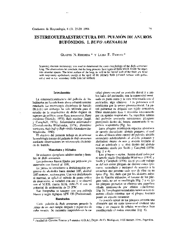
Estereoultraestructura del pulmón de anuros bufónidos. I. Bufo arenarum
ACUEDI
07/02/2015
Descripción
Scanning electron microscopy was used to determined the inner morphology of the Bufo arenarum lung. The observations let conclude that the lung presents three types of folds which divide the organ into alveolar spaces. The inner surface of the lung, as well as the lateral wall of the folds are lined with respiratory epithelium, except in the apex of the primary folds (ciliated surface with goblet cells) and in the secondary folds (ciliated surface).
Hermida, G. & Fiorito, L. (1994). Estereoultraestructura del pulmón de anuros bufónidos. I. Bufo arenarum. Cuadernos de Herpetología, 8 (1), pp. 25-29
Categorias:
Colecciones:
Recuerda
Si te gusta algún autor, colabora con él comprando sus libros.
La cultura y la educación necesitan de tu apoyo activo.
La cultura y la educación necesitan de tu apoyo activo.
Información del autor

ACUEDI
ACUEDI son las siglas de la Asociación por la Cultura y Educación Digital. Somos una asociación civil sin fines de lucro, con sede en Lima (Perú), fundada en noviembre del 2011. Nuestro principal objetivo es incentivar la lectura y la investigación académica, especialmente dentro de espacios digitales. Para ello hemos diseñado una serie de proyectos, todos ellos relacionados entre sí. Este es nuestro proyecto principal, nuestra Biblioteca DIgital ACUEDI que tiene hasta el momento más de 12 mil textos de acceso gratuito. Como tenemos que financiar este proyecto de algún modo, ya que solo contamos con el apoyo constante y desinteresado de la Fundación M.J. Bustamante de la Fuente, hemos creado otros proyectos como ACUEDI Ediciones, donde publicamos libros impresos y digitales, y la Librería ACUEDI, donde vendemos libros nuestros y de editoriales amigas ya sea mediante redes sociales, mediante esta plataforma, en eventos o en ferias de libros.Colabora con
ACUEDI
ACUEDI
